關於長佳
創辦人與企業使命
企業概要
菁英人才管理團隊
合作夥伴
公司里程碑
產品與解決方案
醫療人工智慧
癌症放射治療 AI
急診與放射影像 AI
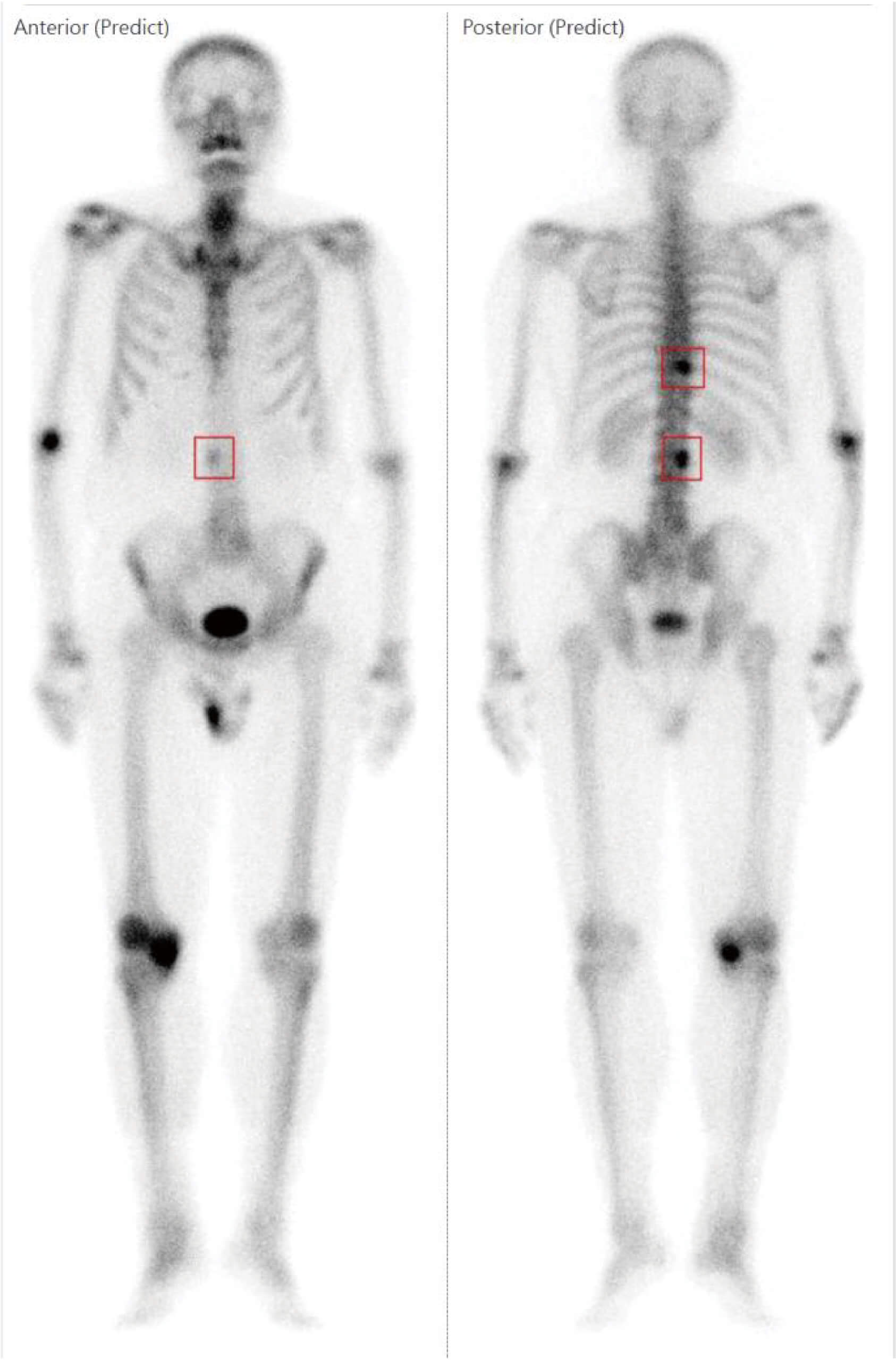
骨閃爍顯像電腦輔助偵測平台
心肌灌流影像分析報告系統
雲端生醫平台
雲端生醫平台總覽
基因檢測結果AI分析平台
智能化健檢排程系統
AI頭皮養護管理平台
AI醫療影像辨識管理平台
生技醫美
芮妮絲極泌奇肌精萃
芮妮絲極泌蘊髮精萃
最新消息
活動消息
公司消息
財務消息
ESG
ESG永續發展首頁
社會共好
永續報告書
供應商永續管理
供應商管理政策
永續供應鏈管理
永續環境
環境管理
溫室氣體管理
水資源及能源管理
廢棄物管理
環保支出
產品品質與客戶服務
投資人與利害關係人
公司治理
組織架構
公司治理相關辦法
公司治理主管
風險管理
智慧財產權管理計畫
人權政策與具體管理方案
內部稽核組織與運作
違反職業道德之申訴管理
禁止內線交易規定
各項員工福利措施
員工工作環境與人身安全
企業誠信經營專職單位運作及落實誠信經營情形
高階經理人薪酬與ESG績效連結政策
資訊安全政策
功能性委員會
審計委員會
薪資報酬委員會
提名及風險管理暨永續發展委員會
董事會
董事資訊
董事簡歷
獨立董事資訊
與獨立董事溝通情形
董事會重要決議事項
董事會多元化資訊
董事會成員及重要管理階層之接班規劃
財務資訊
每月營收
財務報告書
股東專區
致股東報告書
重大訊息公告
主要股東名單
股務資訊
股東會資訊
投資人關係
利害關係人專區
溝通管理
聯絡洽詢資訊
法說會及相關資料
人才招募
招募流程
友善職場
聯絡我們
繁體中文
繁體中文
English
Beyond Medicine.
Anytime. Anywhere.
News
2026-03-13
《AI創新百強》解決醫師人力吃緊!長佳智能用AI重寫放療流程,勾勒時間縮短84%、加快患者治療進度
2026-01-21
衍生企業長佳智能 基因寶銅獲殊榮
2025-12-18
醫療機器人連上AI大腦 中醫大附醫X長聯科技共創「愛寶」量產在即 引領智慧照護新未來
2025-12-18
兒童生長平台與生成式AI照護機器人「愛寶」雙雙獲獎
2025-10-23
醫院自己打造護理機器人!中醫大主導×長聯研發,實現「醫療主權AI」
2025-09-26
攻500億商機 長佳智能推照護機器人
2025-09-26
台灣首款「生成式AI照護型機器人 愛寶 」亮相!
2025-09-19
恭喜!蔡長海 蔡董事長榮獲 工研院產業學院 新科院士殊榮!
2025-09-15
長佳智能奪冠!長聯科技勇奪亞軍
2025-08-19
長佳智能鏡周刊專訪-AI醫療正加速進軍國際!
2025-08-12
長佳智能 × 群聯電子 × 中國醫成立「長聯科技」引爆 AI 醫療機器人新紀元!
2025-05-11
長佳智能醫療照護機器人拚明年底取證 量產無時間表
2025-03-28
長佳智能攻AI照護機器人 攜手群聯成立長聯科技
2025-02-27
從一間區域醫院到生技創新火車頭,揭中國醫孵化3明星生醫股戰法
2025-02-21
長佳智能氣管內管深度檢測系統 獲美國FDA醫材許可
0
取得醫材許可證
0
合作夥伴
0
技術專利
Top-Notch Medical
AI platform
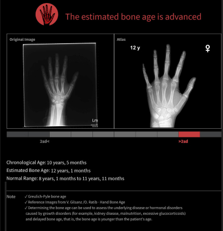
骨齡輔助診斷系統
骨閃爍顯像電腦輔助偵測平台
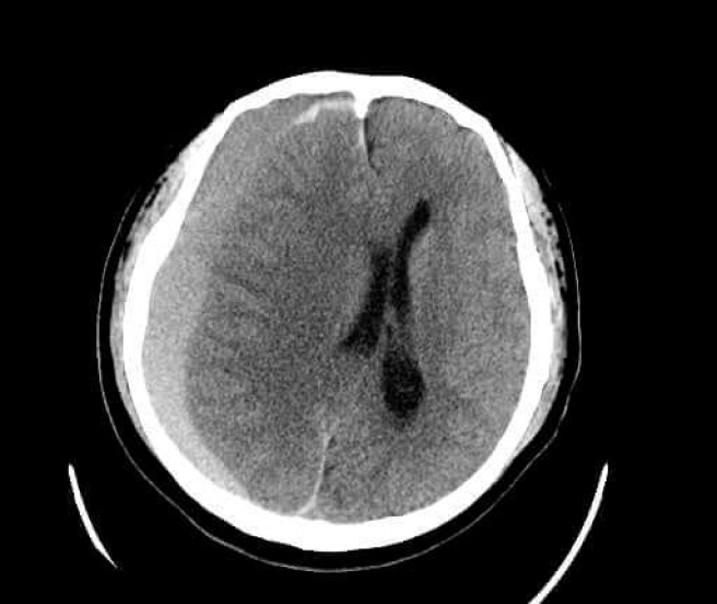
腦出血與腦中線偏移檢測系統
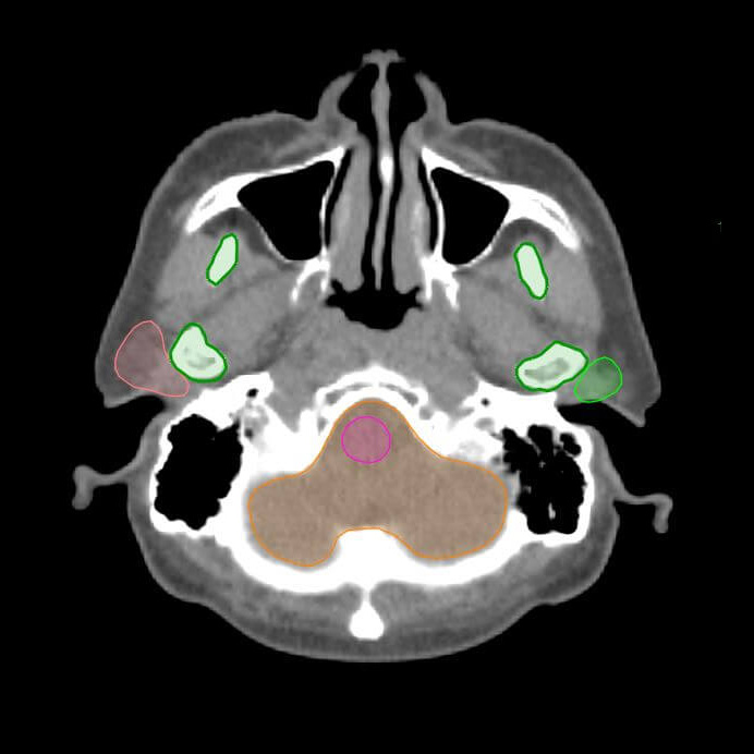
放射治療器官自動勾勒系統
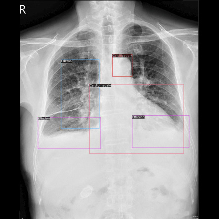
人工智能輔助胸部X光診斷系統
心肌灌流影像分析報告系
產品展示預約
預約展示
您的姓名
服務單位
職稱
*如果沒有所屬服務單位及職稱,請填寫無。
聯絡電話
電子信箱
預約展示的產品
請選擇產品
癌症放射治療 AI
急診與放射影像 AI
骨閃爍顯像電腦輔助偵測平台
心肌灌流影像分析報告系統
基因檢測結果AI分析平台
智能化健檢排程系統
AI頭皮養護管理平台
AI醫療影像辨識管理平台
聯繫內容
清除重填
確認送出
請稍後...
PAGE TOP
