How it works
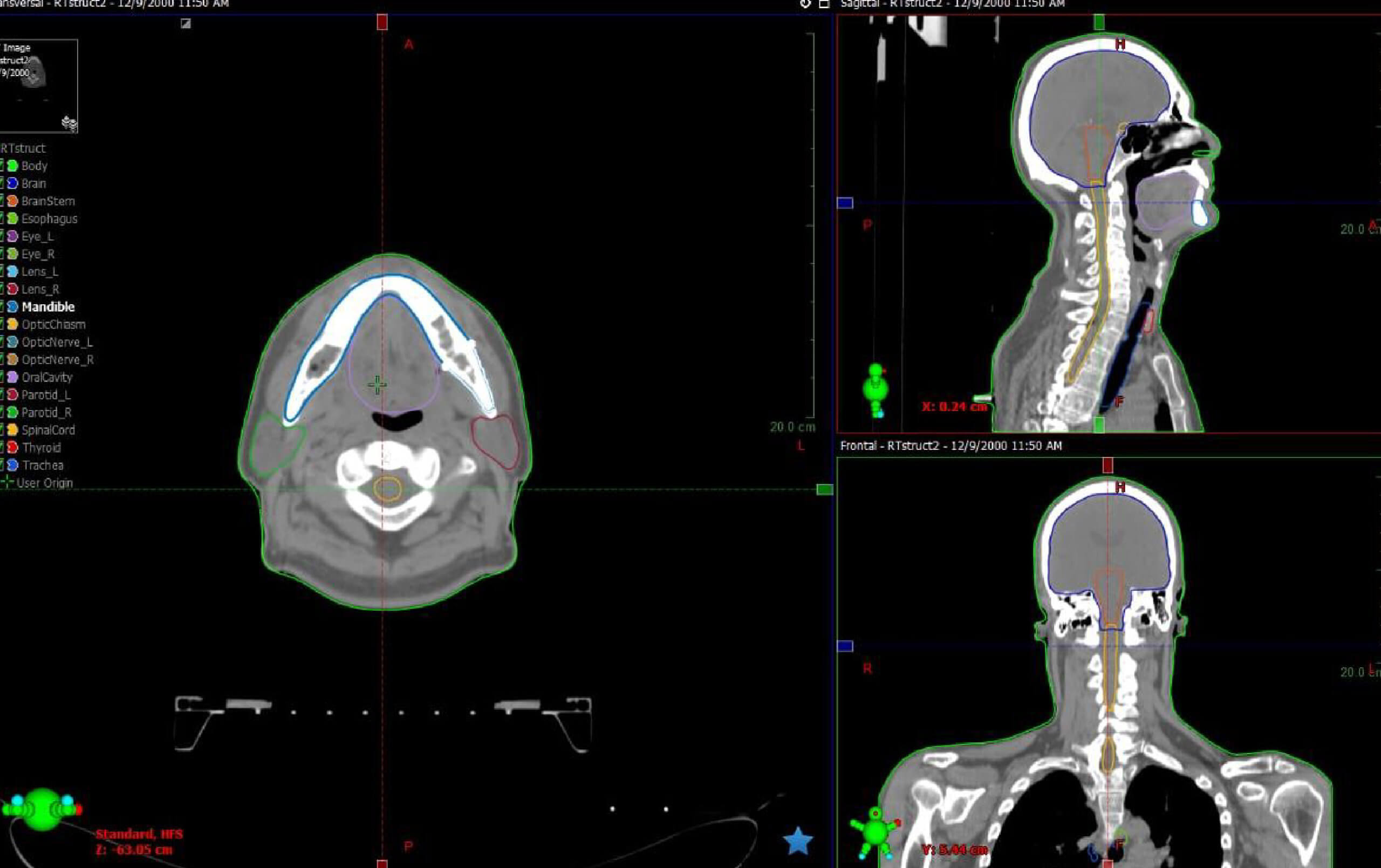
Automatically delineate 17 types of head-and-neck organs-at-risk on CT images and assist radiation oncology professionals in expediting treatment planning workflow
Clinical challenges and unmet need
Over 50% of cancer patients require at least one radiotherapy as part of cancer care 1
Automatically delineate 17 types of head-and-neck organs-at-risk on CT images and assist radiation oncology professionals in expediting treatment planning workflow 2
-
Globocan 2020
*1
-
Trapani D, Murthy SS, Boniol M, et al. Distribution of the workforce involved in cancer care: a systematic review of the literature. ESMO Open. 2021;6(6):100292.
*2
50% of cancer patients
require at least one radiatiotherapy.
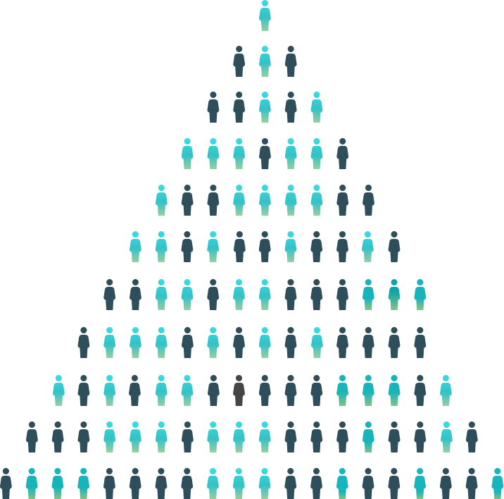
On average, only 0.15 radiation oncologies available per 100 cancer patients

No Data Found
Time saving
0
%
Performace

No Data Found
Pre-EFAI: 4-5 hrs (manual segmentation)
Post-EFAI: 10-15 mins


Previous
Next
17 OARs in head and neck region
Brain
Brain Stem
Eye (left/right)
Lens (left/right)
Optic Chiasm
Brain Stem
Eye (left/right)
Lens (left/right)
Optic Chiasm
Optic Nerve (left/right)
Parotid (left/right)
Oral Cavity
Mandible
Parotid (left/right)
Oral Cavity
Mandible
Spinal Cord
Esophagus
Thyroid
Trachea
Esophagus
Thyroid
Trachea
17
OARs
Key benefit
- Improve efficiency of OARs
Automatic organ segmentation process shortens the time required for contouring
- Standardize segmentation process
Reduce contouring variation among physicians and ensure the consistency and accuracy
- Simplify workflow
Seamless send contoured images to the chosen treatment planning system (TPS) for clinicians to review, edit, and approve. Free up physician's time to focus more on patient care
Explore product

Oncology
RTsuite CT HN-Segmentation System
Want to learn more? Reach out to us
Successful Case
Successful case studies in healthcare, insurance and health and wellness food
Rtsuite Ct Hcap-Segmentation System
Rtsuite Ct Hcap-Segmentation System
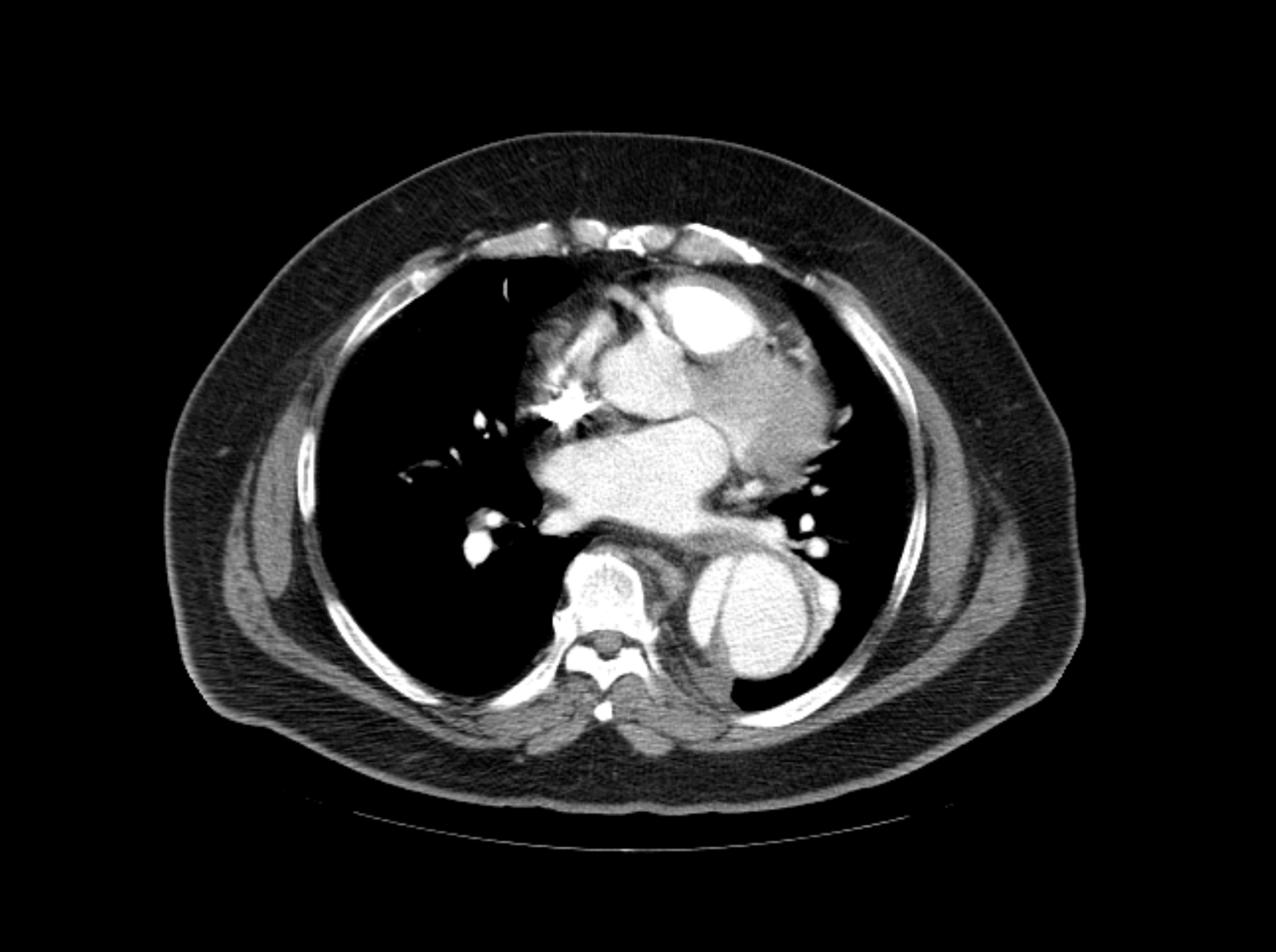
Automatically delineate 80 critical organ structures on computed tomography (CT) images, including regions such as the head, neck, and pelvic lymph nodes. Highly compatible, it works with any brand of treatment planning system available in the market, ensuring seamless integration with various CT and TPS systems
Computer-Assisted Detection Platform for Bone Scintigraphy
Computer-Assisted Detection Platform for Bone Scintigraphy
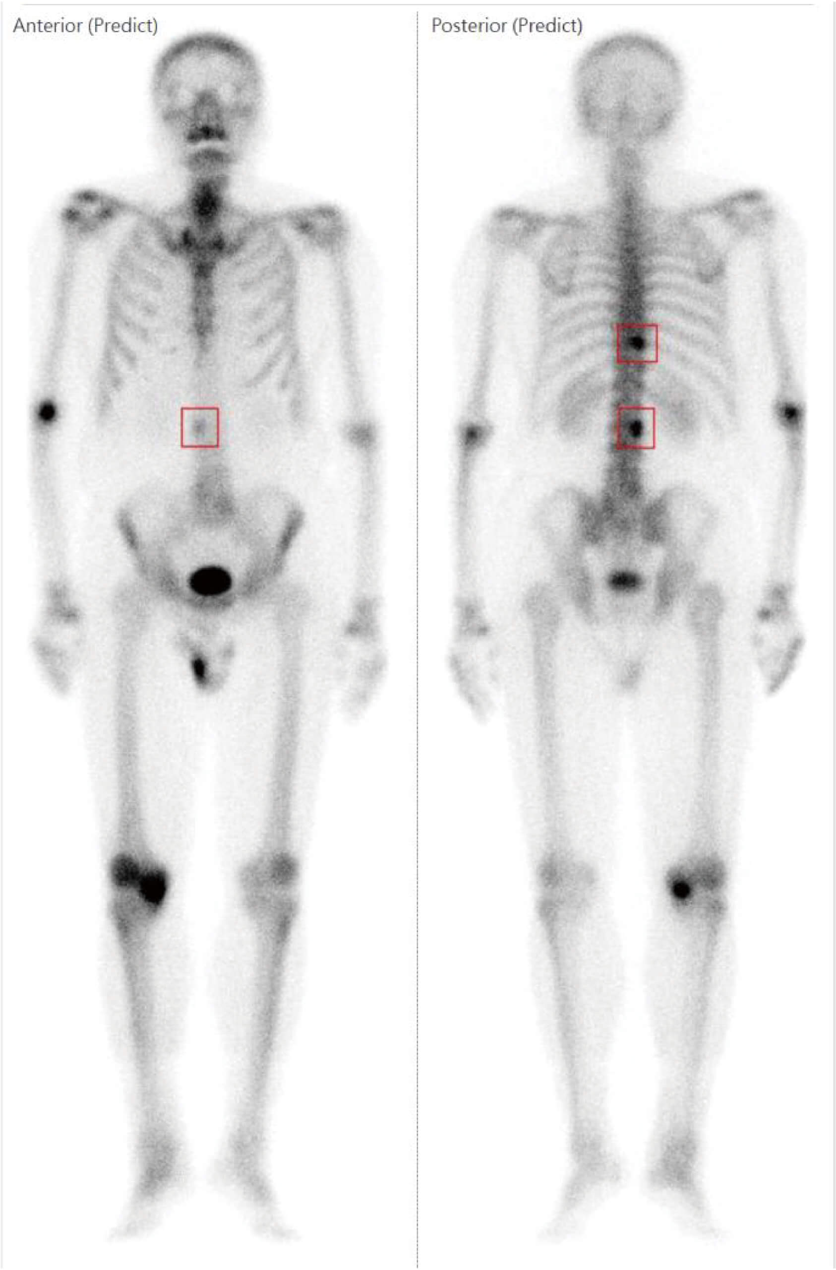
Through artificial intelligence algorithms analyzing whole-body bone scintigraphy images, it assists nuclear medicine specialists in examining the distribution of hotspots. This aids in detecting potential bone metastases, while also highlighting suspected areas for reference by clinical physicians
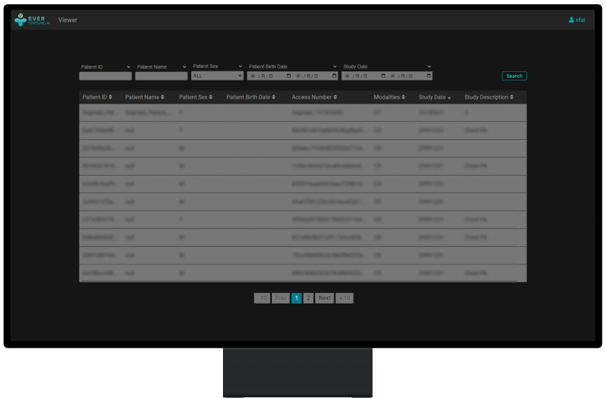
PACS Picture Archiving and Communication System
PACS Picture Archiving and Communication System
A system for managing digital images and communications in medicine. Assists in displaying, processing, storing, and transferring data in compliance with DICOM images. Enable filtering, digital manipulation and quantatitve measurements as well.
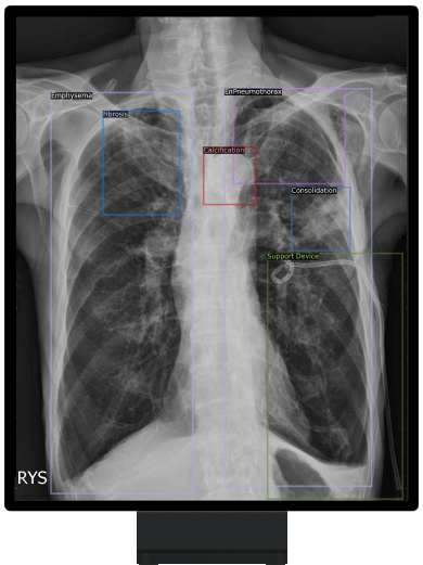
ChestSuite XR Assessment System
ChestSuite XR Assessment System
Identify 15 abnormal finding in chest X-ray images with heart, lungs and bones. The system as a pre-read assistance enable a quick interpretation and faster decisions

RTSuite CT HN-Segmentation System
RTSuite CT HN-Segmentation System
Automatically delineate 17 types of head-and-neck organs-at-risk on CT images and assist radiation oncology professionals in expediting treatment planning workflow
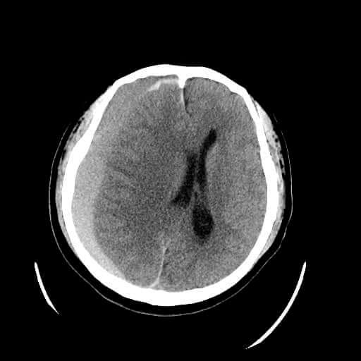
NeuroSuite CT ICH Assessment System
NeuroSuite CT ICH Assessment System
Accurately detect intracerebral hemorrhage (ICH) and identify five types of ICH in non-contrast computed tomography (CT) scan
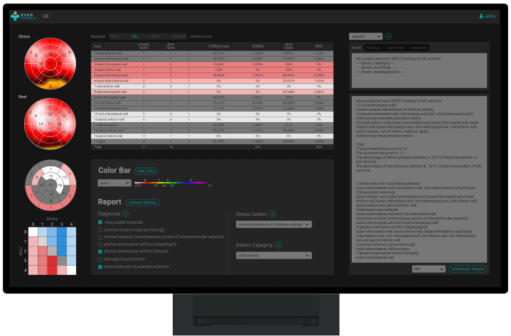
Cardiosuite SPECT Myocardial Perfusion Agile Workflows
Cardiosuite SPECT Myocardial Perfusion Agile Workflows
Provide nuclear radiologists a complete review and quickly modify of paitent's report to shorten the time for diagnostic and clinical decision making.
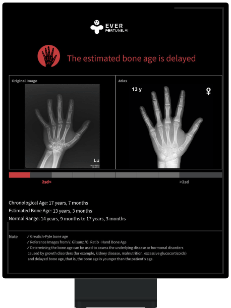
Computer-Aided Bone Age Diagnosis System
Computer-Aided Bone Age Diagnosis System
Evaluate left hand X-ray images of children and adolescents at age 2 to 16 years to assess bone age. Assist pediatricians or clinicians distinguish if a child's bone development is normal, delayed, or advanced for diagnosis and treatment
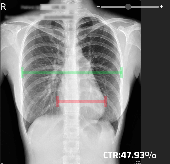
Intelligent Cardiothoracic Ratio(iCTR) Assessment System
Intelligent Cardiothoracic Ratio(iCTR) Assessment System
Automatically measure the maximal transverse diameter of heart and maximal inner transverse diameter of thoracic cavity further to calculate the cardiothoracic ratio of a chest X-ray image. Enable outputting structured reports, optimizing report generation efficiency, and assisting different types of physicians in focusing more on clinical decision-making and patient care.
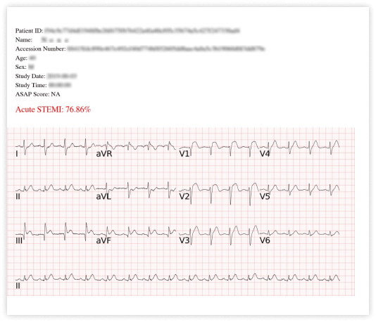
STEMI Detection Software
STEMI Detection Software
Analyze 12-lead electrocardiograms to assist healthcare professionals in quickly detecting acute myocardial infarction, in order to increase the chance of early treatment for patients with acute myocardial infarction
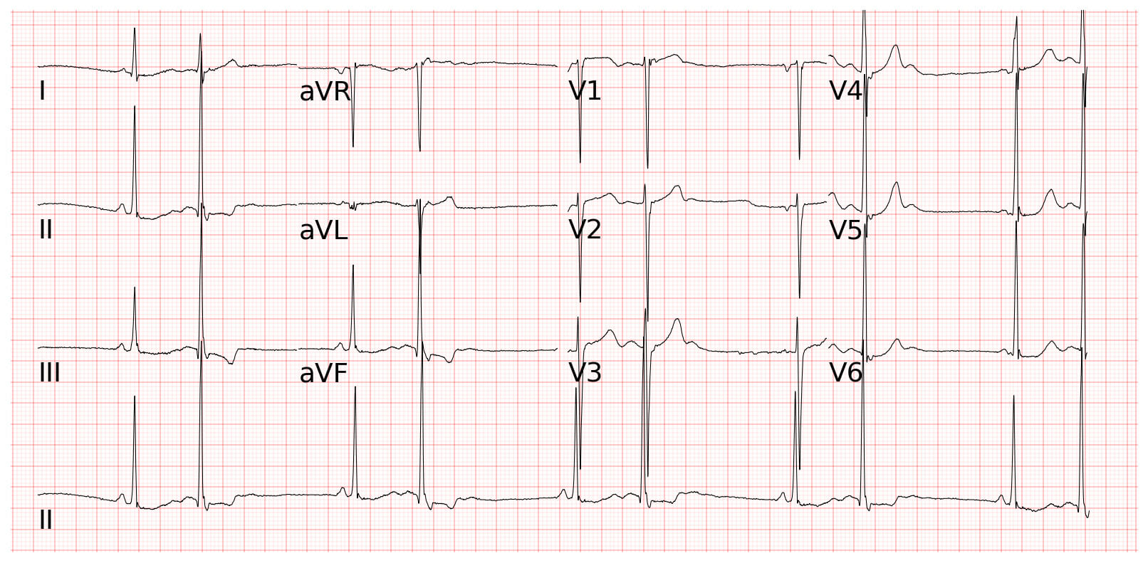
ECG Analysis Software
ECG Analysis Software
Applicable for outpatient and emergency department. Able to analyze rhythm interpretation results automatically from 12-lead electrocardiograms and output the results to assist clinical physicians in quickly identifying heart anomaly and providing appropriate treatment.
prev
next